![]()


![]()


Bioluminescent cell images are illuminated
Bioluminescent cell images are illuminated
10 лютого 2025, 8:49
Imaging living cells with fluorescent proteins has long been a key method for understanding cellular behavior. Although bioluminescent proteins have several advantages over fluorescent proteins, the limited choice of colors has made it difficult to observe multiple targets simultaneously. Now, researchers at SANKEN (Institute of Scientific and Industrial Research) at Osaka University have developed a revolutionary method to expand the color palette of a bioluminescent protein to 20 different colors, enabling advanced simultaneous multicolor imaging.
Cells are the fundamental building blocks of life. Understanding how they function is essential for progress in the life sciences, medicine, and drug discovery. Optical labeling techniques allow scientists to observe cell behavior, track their fate, and identify cells with specific characteristics. Although fluorescent proteins are widely used for these purposes, bioluminescent proteins are gaining popularity due to their unique advantages.
Bioluminescence, the natural emission of light by living organisms, is provided by a chemical reaction catalyzed by an enzyme, usually luciferase, acting on a bioluminescent substrate. Unlike fluorescent proteins, bioluminescent proteins do not require external light for excitation, which avoids problems such as phototoxicity and background light. However, their use is limited by the small number of colors available. Having clear and easily distinguishable colors is vital for simultaneously tracking multiple targets.
Previously, a five-color series of bioluminescent tags was created by combining one of the brightest luciferases, NanoLuc, with a fluorescent protein. This method utilizes the transfer of excited state energy from the substrate to the fluorescent protein, changing the color of the bioluminescence. While this five-color palette was effective, it was not sufficient for more complex imaging needs. Researchers at Osaka University have solved this problem by expanding the bioluminescent color palette to 20, making a significant leap forward in multicolor imaging technology.
“Instead of combining NanoLuc with one fluorescent protein, we combined it with two,” says lead author Mitsuru Hattori. “This approach allowed us to access a much wider range of bioluminescence colors by fine-tuning the combinations of fluorescent proteins.”
The researchers made significant progress with their new bioluminescent protein tags. They have obtained a single-shot image of a mixture of cells expressing all 20 bioluminescent proteins, used the tags to visualize individual subcellular components, and even demonstrated their ability in live mice. In addition, they have successfully performed time-lapse observations of cell behavior over several hours while simultaneously tracking seven different labels.
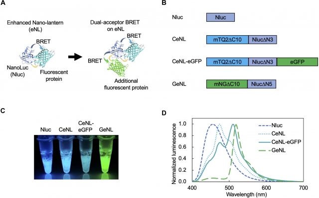
Fig. 1. Spectral changes in eNLs upon introduction of dual-acceptor BRET.
(A) Schematic representation of the spectral changes in the enhanced nanolantern (eNL) with a dual acceptor. (B) Protein structures of luciferases. mTQ2, mTurquoise2; mNG, mNeonGreen. eGFP was attached to the C-terminus of CeNL. (C) Bioluminescence images of purified Nluc and eNL variants obtained with a smartphone camera. Substrates were added to the purified proteins. Exposure time: 0.5 s. (D) Bioluminescence spectra of eNL variants. The intensity is normalized to the corresponding peak values.
“What is truly exciting is that we were able to detect all 20 colors simultaneously, without any time delay, using a standard smartphone camera,” explains senior author Takeharu Nagai. “This innovation makes it much easier and more cost-effective to monitor multiple targets or track individual cells in a population.”
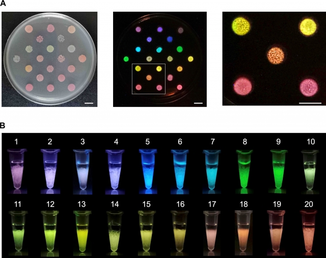
Fig. 2. Twenty color variants of bioluminescent proteins, eNLEX.
(A) E. coli colonies expressing each bioluminescent protein. Shown are the light field image (left), the bioluminescence image (middle), and the magnified white square image (right). Exposure time for bioluminescence: 10 с. Scale bar: 10 mm. (B) Bioluminescence images of purified eNLEX members taken with a smartphone camera. 1: mCRedNL-tdT, 2: mCXL7NL, 3: CeNL-tdT, 4: mCXL2NL, 5: Nluc, 6: CeNL, 7: CeNL-eGFP, 8: GeNL, 9: YeNL, 10: OeNL-mTQ2, 11: OeNL-eGFP, 12: OeNL-Venus, 13: GeNL-tdT, 14: OeNL, 15: OeNL-mKOκ, 16: OeNL-tdT, 17: ReNL-eGFP, 18: ReNL-Venus, 19: ReNL-mKOκ, 20: ReNL. Exposure time: 0,5 с.
These newly developed bioluminescent dyes have the potential to revolutionize cell fate tracking, offering insight into how cells transform into specific cell types and identifying cells with unique drug responses. The team's breakthrough in bioluminescence imaging opens up new possibilities for the development of biological research, drug development and medical science. With such a bright “rainbow” of bioluminescent colors, who knows what scientific treasures lie ahead?
Source: Mitsuru Hattori, Tetsuichi Wazawa, Mariko Orioka, Yuki Hiruta, Takeharu Nagai. Creation of desired bioluminescence colors for simultaneous multicolor biovisualization. Science Advances, 2025; 11 (4) DOI: 10.1126/sciadv.adp4750